EOG
Definition:
Electrooculography (EOG) is a biopotential recording used to examine movements of the eye.
Amplitude:
0.4-1 mV
Frequency:
DC-100 Hz
Typical Applications:
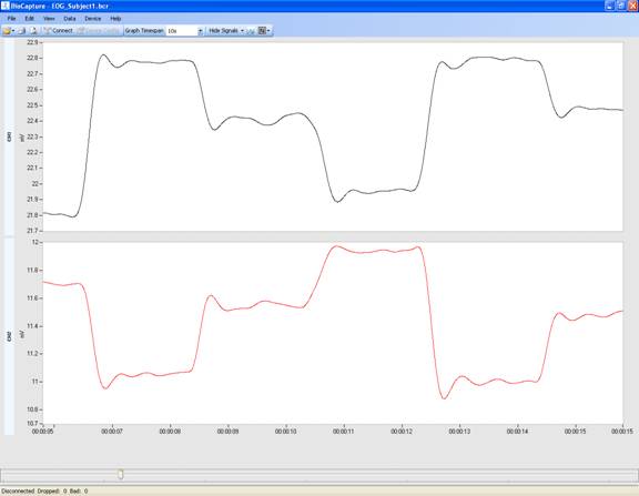
Clinically, the EOG is used to assess eye position and eye movements, especially during sleep studies. It is also used in a variety of psychophysiology research studies.
Typical BioRadio Configuration:
The BioCapture software has a Standard Configuration option that provides a pre-set configuration for EOG.
- Select Device Config and then EOG from the drop down menu
- This will automatically set the typical configuration parameters for EOG:
- Coupling: DC
- Range: ±18 mV
Typical Setup:
- Electrodes: Snap/Cloth
- Adequately prepare the skin before beginning:
Step 1:
Clean the skin on the side of each eye (the area between the eye and the hairline) and in the middle of the subject's forehead with an alcohol padStep 2:
Add a small amount of electrode conducting cream to each electrode- Note: Abrading the skin for an EOG is not recommended.
Step 3:
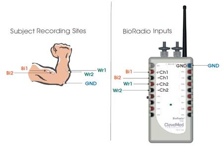
The ground of an EOG should be placed on a bony structure. Typically, the ground is placed on the center of the subject’s forehead- The following image provides an example of common EOG electrode placement:

Common Questions:
Q: I want to examine gross eye movements, such as during sleep, for my EOG study. What coupling should I use?
A:
If you are examining gross eye movements only and do not want to quantify eye position, you should use AC coupling.
Q: I want to examine eye position for my EOG study. What coupling should I use?
A:
If you are examining eye position, you should set your coupling to DC.
Q: Why is my signal not changing and staying constant at approximately 18 mV?
A:
The subject you are collecting data from may have a very large EOG signal, causing your signal to saturate in this preconfigured range. Select Device Config from the menu bar. Under Advanced View, change the range for the EOG channel that is clipping to ±70 mV and program the device.


