EMG
Definition:
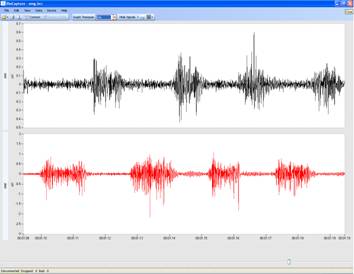
Electromyography (EMG) is a biopotential recording on the surface of the skin used to examine the electrical activity of the muscles.
Amplitude:
: 50 μV-5 mV for skeletal muscle
Frequency:
Skeletal muscle: 2-500 Hz; Smooth muscle: 0.01-1 Hz
Typical Applications:
Clinically, EMG is used to diagnose neuropathies, myopathies, and neuromuscular junction diseases. Experimentally, EMG is used in kinesiology, prosthetics, and as an interface for various control systems, such as prosthetics and computers.
Typical BioRadio Configuration:
The BioCapture software has a Standard Configuration option that provides a pre-set configuration for EMG.
- Select Device Config and then EMG from the drop down menu
- This will automatically set the typical configuration parameters and filter settings for EMG.
- Coupling: AC
- Range: ±25 mV
- Filter: 2nd order, highpass, Butterworth with an upper cutoff of 30 Hz
Typical Setup:
- Electrodes: Snap/Cloth
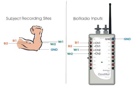
- Electrode Placement: EMG can be recorded from any superficial muscles. As an example, typical arm EMG placement for recording the biceps and wrist extensor muscles is below:

- Ensure that you accurately prepare the skin before placing the electrodes.
Step 1:
Lightly abrade the skin with pumiceStep 2:
Clean skin with an alcohol wipeStep 3:
You may also need to remove any hair that is in the location of the electrode because presence of hair could result in poor electrode/skin contact and contribute to motion artifacts. Step 4:
Palpate the muscle with your finger while you are contracting the muscle so you can locate it. Once you locate the targeted muscle, place two electrodes near the motor point, spaced approximately two inches apart.
Note:
Make sure the skin is dry when you place the electrode. Additionally, if the subject begins to sweat the electrode contact will diminish and you may have to repeat this process in order to ensure sufficient skin/electrode contact.
Step 5:
Place the ground on a bony structure. For example:
- For a leg EMG, the ground could be placed on the knee cap
- For an arm EMG, the ground could be placed on the outside of the elbow
Common Questions:
Q: What muscles can an EMG be recorded from?
A:
Any large, superficial muscles can be used to record EMG. It is important to note that if the desired muscle is located beneath other muscles your EMG signal may have interference from other muscles in close proximity.
Q: How can I reduce the amount of motion artifact in my signal?
A:
The first step to reducing motion artifact is by preparing the skin for electrode placement. See Typical Setup for more information. Other steps to reduce motion artifact include braiding and/or twisting the leads together and taping them to the skin to reduce the amount they move. Any motion on the leads themselves can create artifact. CleveMed also provides an optional artifact removal cable accessory that can help reduce motion artifact. Finally, you could apply a high pass filter at 30 Hz to reduce motion artifacts.


