ECG/ EKG
Definition:
Electrocardiography (ECG/EKG) is a physiological recording used to examine electrical activity of the heart.
Amplitude:
1-10 mV
Frequency:
0.05-100 Hz
Typical Applications:
Clinically, ECGs are used to diagnose abnormal rhythms of the heart, especially those attributed to damaged conductive tissue. Experimentally, ECG can be used to examine heart rate, electrical conduction paths, and exercise physiology response.
Typical BioRadio Configuration:
The BioCapture software has a Standard Configuration option that provides a pre-set configuration for ECG/EKG.
- Select Device Config and then ECG/EKG from the drop down menu
- This will automatically set the typical configuration parameters and filter settings:
- Coupling: AC
- Range: ±12 mV
- Filter: 1st order, Bandpass with a lower cutoff of 0.5 Hz and an upper cutoff of 25 Hz
Typical Setup:
- Electrodes: Snap/Cloth
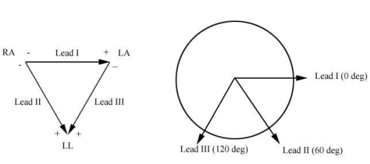
- Electrode Placement: ECG electrodes are commonly placed based on this arrangement where RA is right arm, LA is left arm, LL is left leg, and the right leg (RL) is typically used for ground :

- When placing the electrodes:
Step 1:
Abrade the skin with pumice pasteStep 2:
Clean the area with an alcohol wipeStep 3:
Place electrode cream onto the snap electrode Step 4:
You may also need to remove any hair that is in the location of the electrode because presence of hair could result in poor electrode/skin contact and contribute to motion artifacts
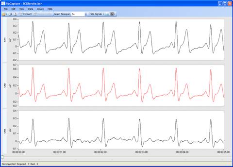
- A normal ECG signal should have a specific wave pattern called the P-wave, QRS complex, and T-wave shown below:

Common Questions:
Q: Why is the signal I am recording opposite of what it should be?
A:
You may have the positive and negative leads reversed.
Q: How can I remove high frequency noise that is appearing in my signal?
A:
One solution to removing high frequency noise is by applying a 60 Hz notch filter. This can be accessed by clicking on the symbol in the taskbar in the software and selecting 60 Hz.
Q: How can I reduce the amount of motion artifact in my signal?
A:
The first step to reducing motion artifact is by preparing the skin for electrode placement. See Typical Setup for more information. Other steps to reduce motion artifact include braiding and/or twisting the leads together and taping them to the skin to reduce the amount they move. Any motion on the leads themselves can create artifact. CleveMed also provides an optional artifact removal cable accessory that can help reduce motion artifact.


